
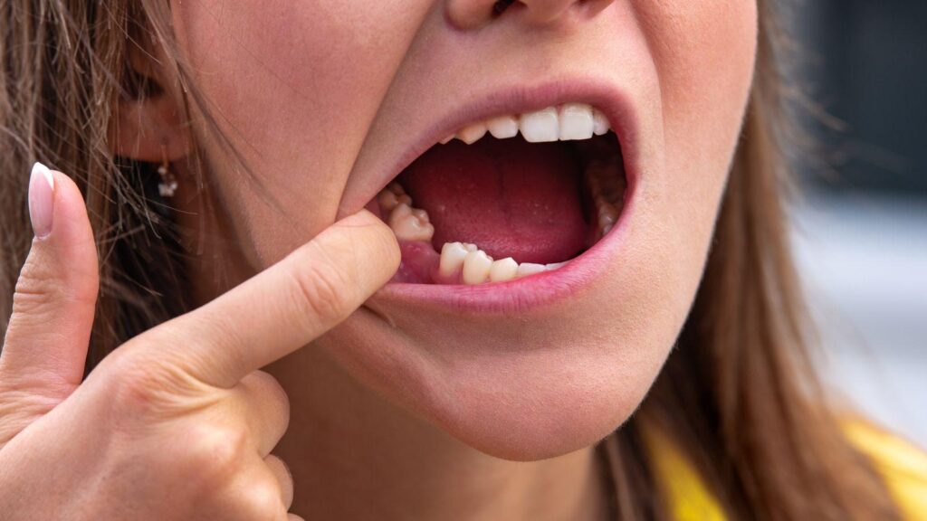
Losing a tooth does more than leave a gap in your smile. The jawbone beneath that missing tooth begins to shrink soon after, and many people don’t notice until rebuilding becomes harder and more expensive.
This article explains what happens inside your jaw and how fast resorption can progress. It also covers common early signs to watch for, who in Millburn faces higher risk, and practical steps you can take right away to limit damage and plan effective repairs with local dental professionals like Arthur Greyf, DDS.
Understanding Bone Structure and Dental Anatomy
Your jaw contains specialized bone that supports teeth and changes when a tooth is lost. Knowing which parts do the work and how they interact helps you understand why preserving bone matters for function and future treatment.
Role of Alveolar Bone in Oral Health
The alveolar bone is the ridge of jawbone that surrounds and holds your tooth sockets. It develops and maintains volume in response to forces transmitted through tooth roots during chewing.
Without those forces, the bone remodels and shrinks. You rely on the alveolar bone for denture support, implant placement, and the contours of your face.
Loss of this bone can change your bite, make dentures unstable, and reduce the available bone for implants, which may require grafting. Key structural features:
- Cortical plate: thin outer layer that provides shape and protection.
- Trabecular bone: porous inner network that absorbs chewing forces.
- Alveolar crest: the highest point of the ridge, important for gum health and implant positioning.
How Teeth Support Jawbone Stability
Teeth transmit mechanical load to the bone via their roots and the periodontal ligament (PDL). The PDL senses pressure and stimulates bone formation where needed, so regular chewing preserves bone density and shape.
When you lose a tooth, the PDL disappears and the localized stimulus for bone maintenance stops. Resorption typically begins within weeks and progresses over months to years, reducing ridge height and width where the tooth once sat.
Clinical consequences you should note:
- Shift in adjacent teeth when support is lost.
- Reduced bone volume that complicates implant placement.
- Altered facial support that can change lip and cheek contours.
Biological Changes Following Tooth Extraction
After a tooth is removed, the local bone and soft tissues begin a predictable cascade of healing and remodeling. You will see immediate changes in the extraction socket and ongoing bone volume loss that can affect surrounding teeth, prosthetic options, and facial support.
Immediate Effects on Surrounding Bone Tissue
When a tooth is extracted, the socket fills with blood clot and then early granulation tissue within days. This tissue stabilizes the site but does not provide the mechanical stimulation the alveolar bone previously received from the tooth root.
Osteoclast activity increases near the socket within the first few weeks, causing resorption of the thin bundle bone that lined the tooth socket. You may not notice outward changes immediately, but the ridge width and height begin to shrink, especially on the facial (outer) side where bone is thinner.
Socket preservation measures taken at the time of extraction—such as grafting with bone substitute and placing a collagen membrane—reduce initial resorption by supporting new bone formation and preserving ridge contours. Primary closure and careful soft-tissue management also limit early collapse of the socket.
Progressive Bone Deterioration Over Time
Most of the measurable bone loss occurs during the first 3–12 months after extraction, with reports of substantial percentage reductions in ridge volume in that period. The process continues thereafter at a lower rate if the site remains unrestored.
Without a root or implant to transmit chewing forces, the alveolar bone undergoes disuse remodeling: osteoclast-mediated resorption outpaces osteoblast formation. This leads to horizontal narrowing and vertical loss of the ridge, which can complicate future implant placement or require ridge augmentation.
Dental implants or implant-supported prostheses counteract this deterioration by providing functional load to the bone. Conventional dentures, however, generally do not stop progressive resorption.
Risk factors that accelerate long-term loss include thin biotype bone, infection at the extraction site, smoking, and systemic conditions like osteoporosis or uncontrolled diabetes.
Warning Signs and Early Indicators of Bone Loss
You may notice changes in comfort, function, or appearance that signal bone loss after a tooth is lost. Timely recognition and imaging are the two ways to confirm and act on those changes.
Physical Symptoms to Watch For
Look for a hollow or sunken feeling where the tooth used to be. The gum contour may flatten or retract, leaving a visible depression that becomes more noticeable over months.
Pay attention if your denture becomes loose or needs frequent relining. Reduced ridge height and width remove the support dentures rely on, causing slipping during chewing or speech.
Notice changes in bite or nearby tooth movement. Teeth adjacent to the gap can tilt or drift, creating crowding, food traps, or difficulty chewing on that side.
Watch for facial changes such as sagging around the mouth or increased wrinkles near the jawline. Those signs reflect loss of underlying bone volume and typically develop gradually.
Detection Through Dental Imaging
Your dentist will use imaging to measure bone loss precisely. Periapical and bitewing X-rays show local bone height and can reveal early resorption around extraction sites or adjacent roots.
Cone beam CT (CBCT) provides three-dimensional assessment of ridge width, height, and density. CBCT is the preferred tool when planning implants or grafts because it shows exact bone volume and anatomical structures.
Compare current images to prior records to track changes over time. Quantifying percent loss or millimeter changes helps determine urgency and the appropriate intervention, such as socket preservation, grafting, or implant timing.
Consequences of Untreated Bone Loss
Untreated bone loss after a tooth extraction can change how you look and how your mouth works. You may not notice the changes at first, but they compound over months and years and affect future treatment options.
Facial Appearance Changes
Bone resorption reduces the height and width of the jaw where the tooth root once stimulated the bone. As bone volume decreases, the soft tissues overlying the jaw lose support; you may see a sunken look around the mouth, deeper nasolabial folds, or a collapsed lower face that shortens your lower facial height.
These shifts can alter how your dentures or partials fit, causing gaps or slippage that make speech and smiling less natural. Bone loss also affects chin projection and cheek contour, so even if other teeth remain healthy, your overall facial proportions can change noticeably over a few years.
Impact on Oral Functionality
Loss of jawbone reduces the stability and support for neighboring teeth, increasing their chance to tilt, drift, or become loose. You may experience new gaps, misaligned bites, or increased wear on the remaining teeth because chewing forces redistribute unevenly.
Reduced bone height limits options for dental implants and can require bone grafting, which adds time, cost, and complexity to restoration. You might also notice chewing becomes less efficient and certain foods feel harder to manage, which can influence nutrition and comfort while eating.
Who Is Most at Risk in Millburn, NJ
Certain local groups face higher chances of jawbone loss after tooth loss. Factors you can control (smoking, dental care) and factors you may not control (age, medical history) both influence how quickly bone resorption progresses.
Prevalence Among Different Age Groups
Older adults in Millburn have a higher incidence of jawbone loss because tooth loss and periodontal disease increase with age. If you are over 60 and have lost one or more teeth, expect a greater rate of bone resorption than someone younger with similar tooth loss.
Younger adults can also be at risk if they lose teeth from trauma or untreated infections. In people under 40, tooth loss is less common, but when it occurs without prompt replacement, local bone loss can still be significant within months to a few years.
Key age-related points:
- 60+ years: higher baseline risk due to cumulative gum disease and past dental work.
- 40–59 years: moderate risk, especially with chronic periodontitis or gaps left unreplaced.
- <40 years: lower population prevalence but rapid local bone changes possible after extraction or injury.
Influence of Pre-Existing Conditions
Certain medical and lifestyle conditions common in Millburn raise your risk for accelerated jawbone loss. If you have uncontrolled diabetes, osteoporosis, or a history of head/neck radiation, your bone remodeling capacity is reduced and resorption after tooth loss accelerates.
Medications and habits also matter. Long-term corticosteroids, some anticonvulsants, and heavy tobacco use impair bone healing and increase resorption.
If you have periodontal disease now, the risk multiplies because active infection already destroys supporting bone. Practical risk flags to discuss with your dentist:
- Diagnosed osteoporosis or low bone density.
- Poorly controlled diabetes (A1c above target).
- Current smoker or heavy vaping.
- Long-term steroid use or recent radiation therapy to the jaw.
Preventative Strategies and Post-Extraction Care
You should focus on protecting the socket, preventing infection, and preserving bone volume when possible. Timely decisions—like immediate grafting or a delayed plan—affect future implant options and the shape of your gums.
Best Practices After Tooth Removal
Follow your dentist’s written post-op instructions exactly for the first 48–72 hours. Bite on gauze as directed to control bleeding, avoid rinsing or spitting forcefully for 24 hours, and sleep with your head elevated to reduce swelling.
Keep the extraction site clean to prevent infection and bone loss. Gently rinse with warm saline or a prescribed antiseptic after 24 hours, and resume brushing surrounding teeth while avoiding direct pressure on the socket.
Discuss socket preservation with your dentist before or at the time of extraction. Options include immediate bone grafting, collagen plugs, or the socket shield technique when clinically appropriate to maintain ridge height for implants.
Manage pain and inflammation with prescribed or recommended OTC medications, ice packs for the first 24 hours, and avoid smoking or using straws for at least one week to prevent dry socket and compromised healing.
Importance of Routine Dental Assessments
Schedule a 1–2 week post-extraction check to confirm soft-tissue healing and to identify early signs of infection or delayed bone repair. Your dentist will assess sutures, mucosal closure, and any persistent tenderness.
Plan follow-up imaging if you intend to replace the tooth with an implant. A CBCT or periapical radiograph at 3–6 months helps determine if additional grafting is needed and guides implant timing.
Keep routine dental cleanings every 3–6 months if you have risk factors like periodontal disease or diabetes. Professional monitoring reduces the chance that adjacent periodontal bone loss will accelerate resorption at the extraction site.
If you notice persistent drainage, increasing pain, or sinking of the gum contour, contact your dental office promptly. Early intervention preserves options for implants, bridges, or other restorative choices.
Modern Solutions for Restoring Lost Bone
You can rebuild jaw volume and create a stable foundation for teeth using targeted surgical techniques and implant options. Treatments range from bone grafts that regenerate local bone to implant types and placements that bypass or stimulate deficient areas.
Bone Grafting Procedures
Bone grafting adds bone or bone-like material where your jaw has resorbed. Your dentist may use autografts (your own bone), allografts (donor bone), xenografts (animal-derived bone), or synthetic materials depending on the size and location of the defect.
Small socket grafts after extraction preserve width and height quickly. Larger defects require block grafts or guided bone regeneration with membranes to protect the graft while new bone forms.
Healing time varies: expect 3–6 months for typical grafts and up to 9–12 months for large reconstructions. Complications are uncommon but can include infection, graft exposure, or partial resorption.
Following post-op care and smoking cessation improves success. Your surgeon will image the site and choose materials and techniques based on bone quality, soft tissue, and your overall health.
Dental Implants as a Restorative Option
Dental implants replace missing teeth and stimulate surrounding bone to slow or stop further resorption. Standard titanium implants require sufficient bone volume for primary stability; when bone is inadequate, implants can follow grafting or use alternative designs.
Options include All-on-4 (angled posterior implants to avoid grafting), zygomatic implants anchored in the cheekbone for severe upper-jaw loss, and short or narrow implants for limited ridges.
Your treatment plan will consider bite forces, number of teeth to replace, and long-term maintenance. Implants typically integrate over 3–6 months, after which restorations attach.
Risks include implant failure and peri-implantitis. Regular hygiene, professional follow-up, and controlled medical conditions reduce those risks.
Collaborating With Local Dental Professionals
You should start by discussing your goals and timeline with a general dentist and an oral surgeon or periodontist. Clear communication helps coordinate extractions, grafting, and any implant planning.
Ask about imaging options such as CBCT scans to evaluate bone volume and anatomy. These images guide whether socket preservation, ridge augmentation, or immediate implant placement suits your case.
Request a written treatment plan that lists steps, costs, and expected healing times. A plan reduces surprises and allows you to compare recommendations from different offices.
Consider a team approach: your restorative dentist, surgeon, and hygienist can each address aspects of function, esthetics, and maintenance. Collaboration improves implant positioning and long-term stability.
Prepare questions before appointments. Useful questions include:
- What bone-preservation options do you recommend and why?
- How soon can I place an implant after extraction?
- What are the risks and expected recovery times?
Verify credentials and experience with bone grafting and implant cases. You want clinicians who document outcomes and use current techniques.
Keep copies of radiographs and operative notes in case you seek a second opinion. Sharing records speeds decision-making and prevents repeated imaging.