
You probably notice differences when you chew or talk, and those everyday forces shape how dental implants in Evansville IN work. The upper jaw has softer bone, closer proximity to the sinus cavity, and different bite dynamics than the lower jaw, so it often requires more implants or additional procedures to achieve the same stability and longevity. Because the maxilla typically offers less dense bone and more surgical constraints, you may need more implants or supplementary treatments (like bone grafts or sinus lifts) in the upper jaw than in the mandible.
This article walks through the anatomical reasons, the functional forces at play, and the prosthetic planning that influence implant number and placement, so you can understand what to expect and why treatment differs between the upper and lower jaws.
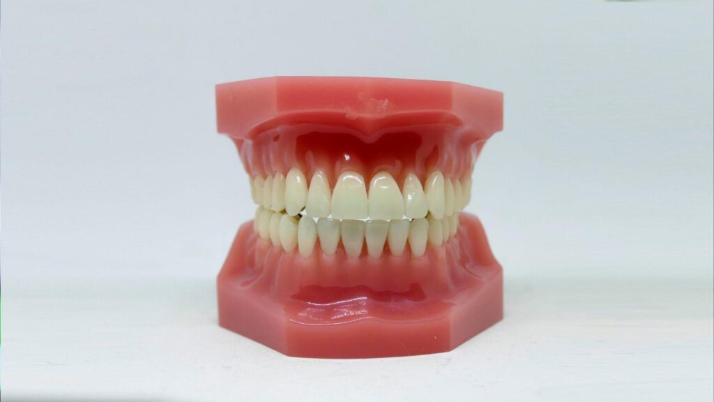
Anatomical Differences Between Jaws
The upper jaw typically has thinner, less dense bone, larger sinuses, and limited vertical height for implants.
The lower jaw usually shows denser cortical bone, a stronger ridge, and the mandibular canal with the inferior alveolar nerve close to implant sites.
Bone Density and Volume Variations
The maxilla (upper jaw) has more cancellous (spongy) bone and thinner cortical plates than the mandible.
You will often find lower bone density in the posterior maxilla, which reduces primary implant stability and increases the need for additional implants or grafting.
The mandible presents thicker cortical bone and greater overall volume, especially in the anterior region.
That denser bone gives implants higher initial stability and often allows you to use fewer fixtures for the same prosthetic support.
When the maxillary sinus expands or pneumatizes after tooth loss, vertical bone height decreases.
You may need sinus lifts or bone grafts to regain platform height for predictable implants.
Implications for Implant Stability
Primary stability depends on bone quality and quantity at the implant site.
In the upper jaw’s softer bone, implants achieve less immediate mechanical stability, raising the risk of micromovement during healing.
You can mitigate that by using more implants, wider or longer implants where anatomy allows, or staged loading protocols.
In the mandible, dense bone often supports immediate or early loading and fewer implants for a given prosthesis.
Osseointegration is slower in low-density bone, so you should allow longer healing periods in the maxilla.
Plan for augmentation or alternative implant designs (tapered, roughened surfaces) when you expect poor bone quality.
Nerve Locations and Sinus Considerations
The inferior alveolar nerve runs inside the mandibular canal, typically 5–15 mm below the alveolar crest depending on jaw height.
You must locate it precisely with CBCT to avoid sensory disturbance when placing lower implants.
The maxilla contains the large maxillary sinus above the posterior teeth.
Sinus proximity limits implant length in the posterior maxilla and often necessitates sinus lifts or short implants.
Nerve and sinus anatomy influence implant number and position.
To protect nerves you may shift implant positions or use angled abutments; to avoid sinuses you may augment bone or choose shorter implants.
Functional Forces Affecting Implant Placement
Functional forces determine how many implants you need, where they go, and how they must be angled and restored. Pay attention to force direction, magnitude, and distribution when planning implant numbers and prosthesis design.
Distribution of Chewing Forces
Chewing forces concentrate differently across the arch. In the posterior maxilla, you typically get higher vertical and lateral loads during mastication, while the anterior region experiences lower, more shear-prone forces. Bone quality in the upper jaw (often thinner, less dense trabecular bone) spreads these loads over less supportive tissue, increasing micromotion risk around implants.
You must account for cantilevers and unilateral chewing habits; both concentrate force on fewer fixtures. Use wider implant distribution, additional implants, or splinted restorations to reduce per-implant load and improve load-sharing.
Impact on Implant Numbers
Higher and less predictable forces in the upper jaw usually force you to plan more implants than for the mandible. Each additional implant reduces load per fixture and lowers the risk of overload-related bone loss. For full-arch maxillary restorations, clinicians commonly place more fixtures or avoid long cantilevers to maintain favorable force distribution.
Patient-specific factors change counts: bruxism, posterior bite dominance, and compromised bone all push implant counts upward. Optimize implant spacing and consider auxiliary support such as zygomatic implants or sinus grafting when standard implants cannot provide adequate distribution.
Role of Occlusal Load
Occlusal load defines long-term implant survival more than implant brand or surface. You need to evaluate maximum bite force, parafunction, and occlusal scheme before finalizing the plan. High occlusal forces create bending moments that concentrate stress at the crestal bone around implants, especially when implant length or diameter is limited.
Control occlusal load through prosthetic design: use mutually protected occlusion, reduce occlusal table width in posterior teeth, and adjust cusp angles. Consider protective measures—nightguards, occlusal guards, or staged loading—to minimize overload during healing and long-term function.
Prosthetic Planning and Treatment Strategies
You will need a plan that balances implant number, position, and prosthetic design to address bone quality, esthetics, and function. Precise planning reduces complications and helps you achieve predictable chewing performance and appearance.
Design of Full-Arch Restorations
Decide between fixed and removable full-arch options based on bone volume, lip support needs, and budget. Fixed hybrid bridges typically require 4–6 implants in the upper arch but often need 6 or more when posterior bone is poor; removable overdentures can work with 4 implants using bar or locator attachments.
Use digital workflows: cone-beam CT for bone mapping, surgical guides for implant angulation, and CAD/CAM milled frameworks for passive fit. Plan prosthetic emergence profiles to preserve papillae and support the upper lip for a natural look.
Account for cantilevers and occlusion. Minimize posterior cantilevers in the upper arch by placing more posterior implants or using zygomatic implants if maxillary posterior bone is absent. Specify occlusal schemes and materials (monolithic zirconia, layered porcelain) to match wear resistance and esthetics.
Support for Dental Prosthetics
Match implant number and distribution to expected functional loads. In the upper jaw, softer posterior bone reduces initial stability, so you may need additional anterior and posterior implants or longer/tilted fixtures to spread forces across bone of better quality.
Choose abutment types and connection designs that reduce micro-movement and screw loosening. Use multi-unit abutments for angulated implants, and consider immediate provisionalization only when insertion torque and implant stability quotient (ISQ) meet thresholds you set.
Plan for bone augmentation when necessary. Sinus lifts, ridge augmentation, or zygomatic/3D-custom implants expand your options but add healing time and surgical complexity. Document load protocol, maintenance intervals, and oral hygiene instructions for long-term prosthetic support.
Long-Term Success Rates
You should expect slightly lower early stability rates for maxillary implants versus mandibular ones due to lower bone density in the posterior maxilla. Long-term survival approaches parity when you address bone deficiency, use proper implant number and distribution, and follow evidence-based loading protocols.
Monitor prosthetic complications separately from biological failure. Framework fractures, veneer chipping, and screw loosening occur more with extensive cantilevers and insufficient implant support; increasing implant count and using stronger materials reduces these risks.
Schedule periodic radiographs and soft-tissue assessments. Track marginal bone levels, ISQ trends, and prosthetic fit at regular intervals to detect issues early and preserve implant and prosthesis longevity.