
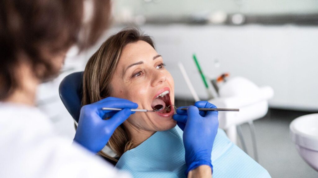
When a toothache won’t quit, you want the cause found and treated fast. Dentists like White Sage Dentistry locate the source quickly by combining a focused clinical exam, targeted imaging, and—when needed—advanced tests or specialist input to pinpoint whether the pain comes from the tooth, the nerve, surrounding tissues, or an entirely different source.
You’ll learn what to expect during the initial assessment. You’ll also see which imaging options reveal hidden problems and when your dentist will use advanced diagnostics or collaborate with specialists.
This article guides you through the practical steps clinicians take. You can feel confident about what happens next and why prompt evaluation matters.
Initial Clinical Assessment
You will gather a focused history, inspect the mouth and surrounding structures, and characterize the pain using specific provocative tests and patient descriptions. Those steps narrow the likely sources quickly and guide which diagnostic tests to order next.
Comprehensive Dental History
Ask when the pain started and how it began; note any recent dental work, trauma, or grinding. Record medical conditions, medications, and allergies that affect inflammation or infection risk (for example, diabetes or anticoagulants).
Document symptom timing and triggers: spontaneous vs. provoked, relation to biting, temperature sensitivity, and whether pain wakes you from sleep. Note any prior similar episodes and previous endodontic or restorative treatments on affected teeth.
Capture social factors that matter: tobacco use, bruxism, and recent sinus or upper respiratory infections. Use a short checklist or form so you don’t miss key items and so you can compare changes on follow-up visits.
Intraoral and Extraoral Examination
Inspect soft tissues, occlusion, restorations, and visible caries under good light and magnification. Look for swelling, sinus tracts, discoloration, mobile teeth, and periodontal pockets that might indicate dental or periodontal sources.
Palpate extraoral lymph nodes and the temporomandibular joints for tenderness, and evaluate facial symmetry to detect nonodontogenic causes like sinusitis or TMJ referral. Percuss suspect teeth gently and compare responses to adjacent teeth.
Use transillumination for cracks and bite-testing devices to reproduce biting pain. Record findings systematically—positive percussion usually suggests periapical inflammation, while localized deep probing suggests periodontal involvement.
Pain Characterization
Have the patient describe pain quality (sharp, throbbing, electric), intensity on a numerical scale, and pattern (intermittent, constant, nocturnal). Specific descriptors narrow causes: sharp, short pain to cold often indicates reversible pulpitis; lingering thermal pain suggests irreversible pulpitis.
Perform targeted tests: brief cold or heat tests on each tooth, and compare responses. Note whether response is immediate, lingering, or absent.
Map referred pain by asking the patient to point to the worst spot and by testing neighboring teeth and sinuses. Combine subjective description and objective test results to prioritize imaging or specialist referral.
Discrepancies between symptoms and findings raise suspicion for non-odontogenic pain. This prompts further ENT or neurologic evaluation.
Diagnostic Imaging Techniques
These imaging methods reveal decay, fractures, infections, and bone changes that physical exams can miss. Each technique balances detail, time, and radiation exposure to help pinpoint the source of your tooth pain.
Digital X-Rays for Early Detection
Digital intraoral X-rays show fine details of tooth structure, root tips, and nearby bone with low radiation exposure. Your dentist will use bitewing X-rays to detect interproximal decay and periapical (PA) views to look for abscesses or bone loss at a tooth’s root.
Images display contrast differences that highlight cavities, recurrent decay under restorations, and the presence of radiolucent lesions indicating infection. Results appear almost instantly on a monitor, which lets your clinician zoom, adjust contrast, and compare to past films for changes over time.
Digital sensors shorten exposure time and make follow-up imaging easier. Expect quick positioning, a momentary bite, and immediate review.
Cone Beam CT Scans
Cone beam computed tomography (CBCT) provides a 3D view of teeth, roots, jawbone, and surrounding anatomy that flat X-rays cannot show. Your dentist will order a CBCT when a complex root fracture, unusual anatomy, or persistent pain with normal 2D X-rays requires more detail.
CBCT slices let clinicians evaluate root canals, detect hairline fractures, locate extra canals, and see the extent of lesions within bone. The scan also clarifies relationships to the sinus or nerve canal—critical when planning treatment to avoid complications.
Radiation from CBCT is higher than single digital X-rays but generally lower than medical CT. Clinicians choose limited-field scans focused on the area of concern to minimize dose.
The scan takes under a minute and provides volumetric data your provider can inspect from multiple angles.
Transillumination Methods
Transillumination uses a focused light source to reveal cracks, internal decay, and marginal gaps without radiation. Your clinician places a bright fiber-optic or LED light against the tooth; cracks and cavities disrupt light transmission and appear as dark lines or shadows.
This method excels for detecting vertical root fractures and early enamel or dentin lesions that may not show on X-rays. It’s noninvasive, quick, and safe for repeated use.
Transillumination complements radiographs and CBCT rather than replacing them. When combined with tactile tests and imaging, it strengthens diagnostic confidence and helps your dentist decide whether restorative, endodontic, or surgical treatment is required.
Advanced Diagnostic Methods
These diagnostic tools target the tooth’s nerve status, the surrounding tissues’ reaction to pressure, and how the tooth responds to temperature changes. Each test gives specific, actionable information that helps localize pain sources quickly.
Pulp Vitality Testing
Pulp vitality testing evaluates whether the nerve and blood supply inside a tooth are functioning. You’ll commonly see two types: electric pulp testing (EPT) and laser or pulse oximetry methods that measure blood flow.
EPT delivers a small electrical stimulus; a reliable response usually means neural conduction is present, but a response does not always guarantee healthy circulation. Interpreting results requires context.
Compare the suspect tooth to adjacent teeth, document response thresholds, and note delayed or exaggerated responses. False negatives occur with immature roots, recent trauma, or heavy restorations; false positives can happen when current spreads to neighboring teeth.
Use vitality testing alongside other diagnostics rather than alone. Record baseline readings, repeat when necessary, and correlate with symptoms, radiographs, and clinical findings.
Percussion and Palpation
Percussion and palpation assess the periodontal ligament and nearby bone for inflammation or infection. Percussion involves tapping the tooth vertically and laterally to detect sensitivity; sharp pain on vertical percussion often indicates apical inflammation.
Palpation checks the soft tissues over the root apices for swelling, fluctuance, or tender induration. Perform tests systematically: compare similar teeth on the opposite side and grade sensitivity (none, mild, moderate, severe).
Note whether pain is localized to one tooth or diffused across multiple teeth, as diffuse pain may point to sinus involvement or referred pain. Combine these findings with radiographs and vitality tests.
Persistent percussion sensitivity with nonvital pulp suggests apical periodontitis. Sensitivity with a vital pulp may suggest occlusal trauma or periodontal issues.
Thermal Testing
Thermal testing uses cold and heat to provoke a pulp response and distinguish reversible from irreversible pulpitis. Apply a cold spray or refrigerant on a cotton pellet for cold testing; for heat, use warmed gutta-percha or a heated instrument.
Record onset time, intensity, and duration of pain after stimulus removal. A short, mild response that quickly subsides indicates normal or reversible pulpitis.
A prolonged, lingering, or severe response—especially if spontaneous pain occurs afterward—suggests irreversible pulpitis. Absence of response can mean necrotic pulp or an unresponsive but still partially vital nerve.
Avoid false readings by isolating the tooth with cotton rolls and dry field, and ensure restorations don’t conduct temperature to adjacent teeth. Always correlate thermal results with clinical signs, percussion findings, and imaging before deciding treatment.
Identifying Non-Dental Causes
Non-dental causes often mimic tooth pain but originate from nerves, sinuses, or jaw muscles. You’ll learn how clinicians test for neuropathic sources and when to seek medical referral for conditions outside dentistry.
Neuropathic Pain Evaluation
You’ll start with a focused history: note sudden, sharp electric shocks, burning sensations, triggers like cold air, and any prior facial surgeries or shingles. Your dentist or pain specialist will perform sensory testing — light touch, pin-prick, temperature — across the painful tooth and adjacent facial zones to map abnormal sensations.
Clinical maneuvers include tapping along the course of the trigeminal nerve branches and trying local anesthetic to see if pain temporarily resolves. If a local block relieves pain, a peripheral source is likely; if not, central sensitization or trigeminal neuralgia becomes more likely.
Imaging (high-resolution MRI) or neurology consultation follows when exam findings suggest neuropathic pain or when symptoms are episodic, lancinating, or resistant to routine dental treatments.
Referral for Medical Assessment
Refer promptly when exam and basic dental tests (percussion, biting tests, radiographs, and pulp vitality) fail to identify a dental cause. Provide the medical team with a concise summary: symptom onset, character, distribution, prior dental work, and response to local anesthetic or analgesics.
Indicate specific concerns on the referral: sinus disease (CT or sinus endoscopy), temporomandibular disorder (TMJ specialist or MRI of TMJ), and neuropathic disorders (neurologist for MRI brain and trigeminal pathway assessment). Include any red flags—progressive neurological signs, facial weakness, or systemic symptoms—so they prioritize imaging and possible neurosurgical or ENT input.
Differentiating Common Tooth Pain Sources
You need precise clues from your symptoms, exam findings, and tests to pinpoint whether pain comes from the tooth surface, the root, or supporting tissues. Focused questions and targeted tests usually separate decay, cracks, pulp inflammation, and periodontal causes quickly.
Distinguishing Between Decay and Fractures
Decay usually causes localized sensitivity to cold or sweets and often shows a visible cavitation or shadow on bitewing radiographs. You may feel sharp, transient pain with a cold stimulus that disappears when removed; this suggests dentin hypersensitivity or early caries.
If pain lingers after cold or becomes severe and spontaneous, suspect pulpal involvement from deep decay. Fractures frequently produce sharp, biting-related pain that comes and goes.
You might report pain when chewing that stops once pressure releases; that pattern points to a cracked tooth. Dentists will use selective biting tests (e.g., Tooth Slooth), percussion, and transillumination to reveal fracture lines.
Bitewing or periapical radiographs can miss vertical cracks, so expect additional imaging like CBCT when clinical suspicion remains high.
Key distinguishing features:
- Decay: sensitivity to sweets/cold; visible lesion; radiographic caries.
- Fracture: bite-triggered sharp pain; positive biting test; transillumination or CBCT may show crack.
Periodontal Versus Endodontic Pain
Periodontal pain often stems from inflamed gingiva, deep pockets, or a draining sinus tract and tends to be localized to the gum and supporting bone. You may notice bleeding on probing, increased pocket depth, tooth mobility, or pus drainage.
Pain usually increases with biting if occlusal trauma or furcation involvement exists. Endodontic (pulpal) pain typically produces spontaneous, severe, or lingering pain that can radiate and disturb sleep.
You might experience prolonged sensitivity to cold and heat or throbbing pain that worsens when lying down. Tests that help differentiate include:
- Percussion: positive in periapical involvement of endodontic origin.
- Cold testing and electric pulp testing: inconsistent or absent response suggests necrosis.
- Periodontal probing: deep isolated pocket suggests endodontic-periodontal combined lesions.
Use both periodontal charting and pulp vitality tests together. A sinus tract tracing with a gutta-percha cone on radiograph helps locate whether the source tracks to the root apex (endodontic) or to lateral periodontal sources.
Collaborative Decision-Making
You will share decisions with your dentist using clear information about symptoms, test results, and treatment trade-offs. Expect structured options, the reasons behind each, and next steps tailored to diagnostic uncertainty and your priorities.
Patient Communication Strategies
Start by listing symptoms with timing, triggers, and what relieves the pain. Give exact examples: “sharp pain during cold exposure for 10 seconds” or “dull ache waking me at 3 a.m.”
This helps your dentist prioritize tests and interpret responses to thermal or percussion exams. Ask for plain-language explanations of each diagnostic test and what a positive or negative result would mean for treatment.
Request to see images (radiographs or CBCT slices) and have the clinician point out suspected areas. State your treatment goals and constraints—cost limits, appointment availability, tolerance for procedures—so the dentist can recommend realistic, staged options.
Confirm the recommended next step in writing or by summary before leaving.
Specialist Referrals
Expect referral when findings are ambiguous, when endodontic or periodontal disease appears likely, or when advanced imaging is needed. Your dentist should explain the specific reason: for example, “CBCT recommended to evaluate a suspected root fracture” or “endodontist for complex root canal anatomy.”
Ask about the referral timeline and whether interim pain control or temporary measures (splint, medicated dressing, analgesics) are appropriate. Request that imaging and clinical notes be sent to the specialist beforehand to avoid repeat tests.
Clarify who coordinates follow-up care and how treatment responsibilities shift after the referral. Confirm whether the specialist will perform definitive therapy or recommend return to your general dentist for restoration or periodontal management.
Timely Follow-Up and Reassessment
You should schedule follow-up visits soon after initial treatment or diagnostic testing to confirm whether pain is resolving. Early reassessment helps catch persistent infection, missed fractures, or referred pain that didn’t show on first exams.
During follow-up, the dentist will repeat targeted tests—thermal, percussion, palpation—and compare symptoms to your initial report. Changes over time, such as improvement or new signs, guide the next steps more reliably than any single visit.
If symptoms persist or worsen, expect additional imaging such as a different-angle radiograph or cone-beam CT to reveal issues missed earlier. Sometimes selective anesthesia or bite tests during reassessment pinpoint a tooth causing referred pain.
Keep a clear symptom log between visits. Note pain intensity, triggers, timing, and any swelling or fever.
This short record gives your clinician objective data and speeds diagnostic decisions. Your practitioner may provide temporary measures—NSAIDs, short-term antibiotics when indicated, or protective splints—while planning definitive care.
These measures reduce acute discomfort but don’t replace reassessment to confirm the underlying cause. If you miss a scheduled follow-up, contact the office promptly.
Failure to return after urgent care can jeopardize treatment outcomes and increase the risk of complications.